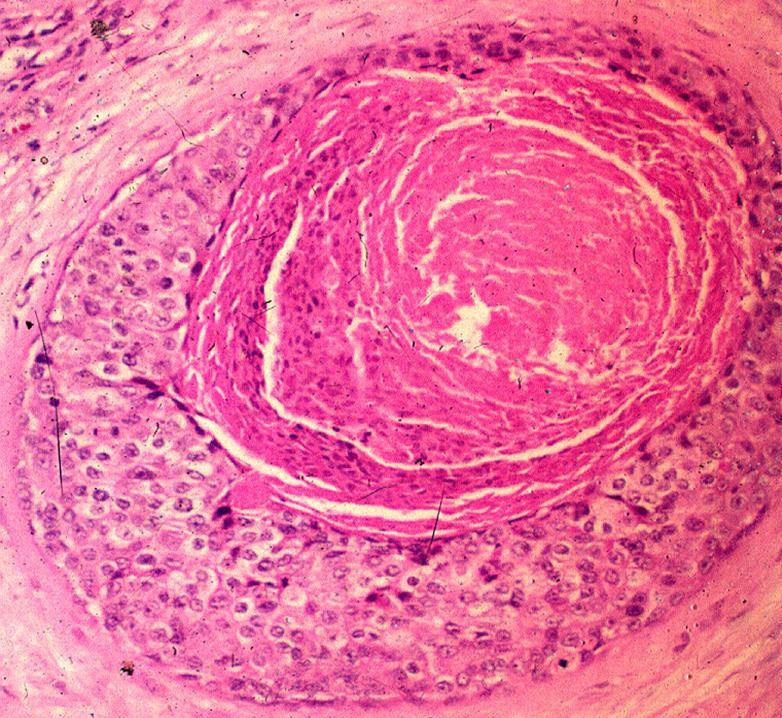
ΤΟ ΜΗ ΔΙΗΘΗΤΙΚΟ ΠΟΡΟΓΕΝΕΣ ΚΑΡΚΙΝΩΜΑ ΤΟΥ ΜΑΣΤΟΥ ΘΕΩΡΕΙΤΑΙ Η "ΚΑΛΥΤΕΡΗ " ΜΟΡΦΗ ΚΑΡΚΙΝΟΥ ΤΟΥ ΜΑΣΤΟΥ

Epidemiology
In the United States, the incidence of DCIS is now 10-20 per 100.000 women-year, with the ratio of DCIS to LCIS being 6/1. The frequency of family history of breast cancer among first degree relatives is not different in patients with DCIS and invasive breast cancer.
Histologic subtypes
DCIS is generally classified as one of five subtypes based on the differences in the architectural pattern and nuclear features. The subtypes are: comedo, solid, cribriform, micropapillary, and papillary. The high nuclear grade and the comedo necrosis are predictive of local reccurence.
Multifocality
Multifocality is generally considered to be present when separate foci of DCIS occure more than 5mm apart in the same breast quadrant. However some investigators believe that multifocal disease may represent the intraductal spread from a single foci of DCIS.
Multicentricity
Multicentricity is difined as a separate focus of DCIS outside the index quadrant. The multicentricity of DCIS is more likely arount 30-40%.
Microinvasion
DCIS with microinvasion is defined as a predominanlty noninvasive disease with foci of invasive cancer less than 1 mm in diameter.
Diagnosis
Microcalcifications are the most common mammograpfic manifestation of DCIS and account for 80% of all carcinoma representing with calcifications. The type with high nuclear grade and comedo necrosis gives linear branchings, on the other hand granular manifestation is associated with micropapillary and cribriform lesions.
The lesions are detected by fine needle aspiration or excisional biopsy and the margins should be at least 1cm, with 2cm being preferable.
Treatment
It is currently recommended that mastectomy be performed for large lesions (more than 3cm) lesions with high nuclear grade or pathologic evidence of close or involved margins. Local excision alone may be appropriated for a select subgroup patients with small and low grade DCIS with free margins and who cab be followed diligently for recurrence.
Local recurrence in the lumpectomy plus radiotherapy is 7% versus 16% in the lumpectomy alone. Radiotherapy appears to decrease the incidence of local recurrence and local invasive recurrence. Patients who would be considered for local excision alone have small tumor (<1cm) low grade and clear margins.
At present tamoxifen should be used in patients with DCID olny in the contect of a clinical trial.
Surveillance
Following breast conservative surgery a post surgical mammogram should be obtain foe residual microcalsification. In addition a mammogram should be obtain 3-4 months after the completion of radiotherapy as a new base line. Follow up of patients after conservative surgery with or without radiotherapy involves annual mammogram and twice clinical examination for 5 years and annual thereafter. The risk of a new primary breast cancer in the contralateral breast after treatment for DCIS approaches two to five times the risk of a first primary breast cancer.
